業內快訊
一個細胞一個細胞地跟蹤早期心臟形成
 時間:2021-01-14
 來源:
來源:
 瀏覽量:3204
近日,研究人員在單細胞分辨率上繪制出了小鼠胚胎心臟起源的圖譜,幫助確定了在發育初期構成心臟的細胞類型。相關論文[Characterization of a common progenitor pool of the epicardium and myocardium]刊登于《科學》。
The mammalian heart is derived from multiple cell lineages; however, our understanding of when and how the diverse cardiac cell types arise is limited. We mapped the origin of the embryonic mouse heart at single-cell resolution using a combination of transcriptomic, imaging, and genetic lineage labeling approaches. This provided a transcriptional and anatomic definition of cardiac progenitor types. Furthermore, it revealed a cardiac progenitor pool that is anatomically and transcriptionally distinct from currently known cardiac progenitors. Besides contributing to cardiomyocytes, these cells also represent the earliest progenitor of the epicardium, a source of trophic factors and cells during cardiac development and injury. This study provides detailed insights into the formation of early cardiac cell types, with particular relevance to the development of cell-based cardiac regenerative therapies.
研究人員對胚胎小鼠心臟進行了顯微解剖,以觀察一種非常早期的細胞條紋——心臟新月,如何轉變為線狀心臟管。研究人員利用高分辨率成像、延時顯微鏡和單細胞RNA測序識別了細胞類型,能夠在大約12小時的發育過程中跟蹤不同群體的心肌祖細胞的發育。
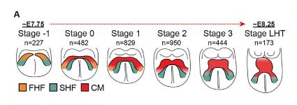
FHF,第一心臟領域;SHF,第二心臟區域;CM,心肌細胞

使用Mab21l2-iCreERT2轉基因小鼠品系進行譜系標記實驗的實驗設計。
示意圖右側突出顯示了在E7.75和E8.0之間標記近心臟區域(JCF)導致在E10.5處標記心肌細胞(CM)和心外膜(Epi)。
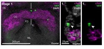
1期Mab21l2-iCreERT2的最大強度投影(MIP)
對心臟肌鈣蛋白T(cTnT)和YFP免疫染色的R26R-YFP胚胎。YFP陽性細胞(箭頭)位于JCF中。
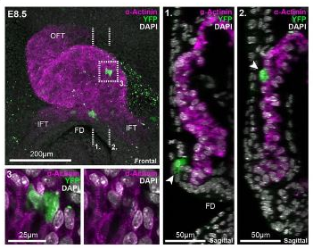
來自E8.5 Mab21l2-iCreERT2的心臟環管的MIP
對α-肌動蛋白和YFP進行了免疫染色。箭頭突出顯示了重組的YFP細胞在心肌內的位置(2.),并保留在JCF祖細胞區域(1.)內。虛線框(3.)放大顯示了JCF衍生的具有肌動蛋白肌節條紋的心肌細胞。OFT,流出道;IFT,流入管道。
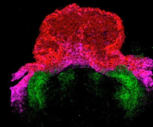
小鼠胚胎心臟初期圖像 圖片來源:牛津大學
圖譜展示了一個老鼠胚胎心臟剛剛開始形成的階段。在初期,這些細胞排列成典型的新月形,并且已經表現出緩慢有節奏的收縮。隨著發育,圖中綠色、紅色和品紅代表了構成心臟的不同區域。胚胎期的心臟在這個階段被描述為“線形心臟管”,并可以看到心室開始形成和成形。
新技術使他們第一次確定了一組祖細胞,它有助于心肌細胞和早期心外膜(心臟最外層)形成。這些膜提供了能指導心臟組織發育和修復的細胞和其他蛋白質。研究人員認為更好地了解它的起源有助于為再生心臟治療提供信息,也可以提高我們對先天性心臟缺陷的認識。
相關論文信息:http://dx.doi.org/10.1126/science.abb2986


